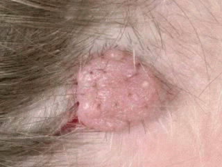
Nevi or moles are defined as benign skin tumors. Histologically they are composed of melanocyte-derived nevus cells. Melanocytes are derived from neural crest tissue. According to age of onset and the location and arrangement of nevus cells within the skin, melanocytic nevi are classified into different types. Melanocytic nevi are arranged as organized clusters of nevus cells located at various levels in the skin. Nevi are equally common among males and females. The average number in adults is between 12 and 20 nevi. In some families a larger number of nevi may be encountered. Irritation by clothing or external trauma may be associated with the development of inflammatory symptoms.
Congenital Nevi
Nevi present at birth or that appear during infancy are termed congenital nevi.
Acquired Nevi
1- The appearance of newly acquired nevi reaches a peak during adolescence.
2- Fewer nevi are acquired after age 30, Nevi appearing after age 30 should be regarded as suspicious.
3- Sun exposure appears to be a stimulus for cell growth of nevi as most acquired nevi appear on sun-exposed skin. Acquired Nevi appearing appearing in sun-protected areas should also be regarded as suspicious.
4- Existing nevi may increase in size and become more heavily pigmented during puberty or during pregnancy.
Development of Nevi
Acquired nevi first appear as flat, round, uniformly colored papules. During this growth phase, nevi expand laterally while remaining flat and symmetric. Nevi may be slightly darker in color and slightly raised in the center, and may remain stable in size and appearance for several years. Over many years, nevi continue to become more elevated and uniformly lighter in color. Eventually the nevus appears as a skin-colored papule or may completely disappear in older years. Residual nevi are rare after age 70.


Sunday, June 22, 2008
 Melanocytic Nevi Pictures
Subscribe to:
Post Comments (Atom)
0 comments:
Post a Comment